transformations
A new generation of tools is enabling scientists to reveal, in breathtaking detail, once-hidden patterns of life.
“From the scientific point of view, images like these are a treasure trove of information,” says Terri Bruce, manager of the Jordan Hall Imaging Facility. “There is a vast amount of detailed data within a single micrographic image.”
Researchers skilled in reading the images can observe, for instance, how an organism develops, the movement of living cells, the arrangements of proteins, or the surface properties of materials.
Today, a picture is worth more than a thousand words. It is worth reams of data.
But the value of images goes deeper than data, Bruce says. She points, for example, to the work of Poulomi Ray, a Ph.D. student in Susan Chapman’s lab in the Department of Biological Sciences.
“One look at the remarkable intricacies of her tissue sections transforms the perceptions of the viewer,” Bruce says. “She makes us see how lovely our world truly is.”
Terri Bruce is a research assistant professor in biological sciences.
life most vulnerable
Susan Chapman’s lab studies how complex organs develop in embryos, a step toward learning how to prevent birth defects. With images like the one to the right, a confocal micrograph of a section of a chick intestine, Chapman and Poulomi Ray can study cells and tissues as they develop.
A similar approach helps Chapman investigate the possibility of regenerating damaged hair cells in the inner ear.
In this research she uses zebrafish, which, unlike people, can naturally regenerate the damaged cells. The image above shows zebrafish stereocilia, which transform the energy of sound waves into electrical signals. Mutations, noise, and chemical exposure can break the links between stereocilia and hair cells.
Chapman’s research studies ways to regenerate these links and restore hearing. The research could lead to therapies for reversing hearing loss in people.
Susan C. Chapman is an assistant professor in the Department of Biological Sciences, College of Agriculture, Forestry, and Life Sciences. Her research is funded by the National Institutes of Health, National Institute on Deafness and Other Communication Disorders.
With powerful new imaging systems, scientists can look to nature for ideas about how to design better instruments and machines. Matt Lehnert, a postdoctoral fellow in entomology, works with a team of engineers and biologists to develop a new device for use in microfluidics, the precise management of small amounts of liquid. Uses for microfluidic devices range from inkjet printers to lab-on-a-chip technologies in medicine. As it turns out, the feeding tube of a butterfly, called the proboscis, is a marvel of microfluidic design.
“The butterfly proboscis previously has been assumed to function only as a drinking straw,” Lehnert says, “but the butterfly proboscis is much more complex than that.”
The research team’s experiments have shown that the proboscis has structures and properties that combine sponge-like capillary action with straw-like sucking. Working together, these structures enable butterflies to take up minute amounts of liquid.
The image to the left shows the proboscis of a red-spotted purple butterfly, Limenitis arthemis astyanax, which feeds on juice from rotting fruit, sap, and sometimes nectar. The scale-like structures along the proboscis are sensory devices.
By studying this system, the research team can learn how very tiny structures take up minute amounts of fluid. This will yield new knowledge about the evolutionary biology of butterflies and other fluid-feeders; it may also lead to microfluidic devices with wide applications, Lehnert says.
Matthew S. Lehnert is a member of a research team led by Konstantin G. Kornev, associate professor of materials science in the College of Engineering and Science, and Peter H. Adler, professor of entomology in the School of Agriculture, Forestry, and Life Sciences.
The National Science Foundation funds this research.
 A tiny sensor imaged by Terri Bruce on the Ti Eclipse confocal microscope.
lab on a chip
A laboratory the size of a computer chip? Yes. Scientists are learning how to create miniature labs that can help analyze tiny amounts of material, even single cells.
Yang Yang, a visiting Ph.D. student from China, designed and fabricated the radio-frequency (RF) sensor above at Oak Ridge National Lab. This ultrasensitive sensor allows scientists to detect and analyze the cell intact, without cutting or labeling. A tube-like structure on the device will enable detection with radio-frequency waves, and the spoke-like AC electrodes are designed to rotate the cell. The RF techniques are expected to be portable, low-cost, and easy to use.
Pingshan Wang, associate professor of electrical and computer engineering, leads the group.
Primary funding is from the National Science Foundation.
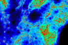
 Tungsten film imaged by Terri Bruce on the Ti Eclipse C1si confocal microscope.
breaking the wave
Flexible electronics (think bendable TV screens) are exciting new devices. One challenge: the bond between the metal circuitry and the polymer backing must be durable.
As part of a collaboration with Neville Moody of Sandia National Laboratory, students in Molly Kennedy’s group studied tungsten films applied to substrates made of PMMA, a polymer. In the upper right hand corner of the image at right, “river marks,” formed as the film initially buckled, led to the kind of separation shown in the lower left.
Ashley Sachs, an undergraduate student in Kennedy’s group, prepared the sample for imaging.
Molly Kennedy is an assistant professor in the School of Materials Science and Engineering, College of Engineering and Science.
Funding for this work is from Sandia National Laboratory.